
Summary
Take-home message. CellMethMarker custom marker regions can recover expected cell-type-specific hypomethylation patterns in an independent human single-cell methylome atlas, supporting their use for annotating unknown methylation samples or clusters.
Background
Single-cell methylation datasets are commonly interpreted by first clustering cells or pseudobulk profiles and then examining cell-type-specific hypomethylated genes or marker regions. CellMethMarker provides a curated reference of cell/tissue-specific DNA methylation markers that can help users assign biological identities to unknown methylation-derived clusters.
- Reference dataset. We used the human single-cell methylome atlas from Zhou, J. et al., which profiled 16 human tissues using the single-nucleus methyl-3C-seq assay. Reference: Zhou, J. et al. Human Body Single-Cell Atlas of 3D Genome Organization and DNA Methylation. bioRxiv. DOI: 10.1101/2025.03.23.644697 (2025).
- Independent validation panel. The atlas contains 86,689 single nuclei and reports 35 major cell types and 206 cell subtypes, providing a suitable external reference for testing whether CellMethMarker markers recover expected cell-type-specific methylation patterns.
- Biological rationale. In methylation-based single-cell or pseudobulk analysis, unknown samples or clusters are often interpreted by identifying cell-type-specific hypomethylated genes or marker regions.
- Objective. We evaluated whether CellMethMarker custom marker regions show expected low methylation in matched Zhou major cell types, and compared this pattern with promoter methylation of representative marker genes from CellMarker2.0.
Methods
We tested whether CellMethMarker marker regions show expected low methylation in matched Zhou major cell types. As a comparison, we also evaluated promoter methylation of representative marker genes from CellMarker2.0.
- Reference tracks: downloaded bigWig methylation profiles for 35 Zhou major cell types.
- Marker sets: evaluated CellMethMarker RRBS and WGBS custom markers separately.
- mCG extraction: extracted mCG fractions at marker regions across all reference cell types.
- Heatmap analysis: visualized raw methylation and row-wise z-score patterns.
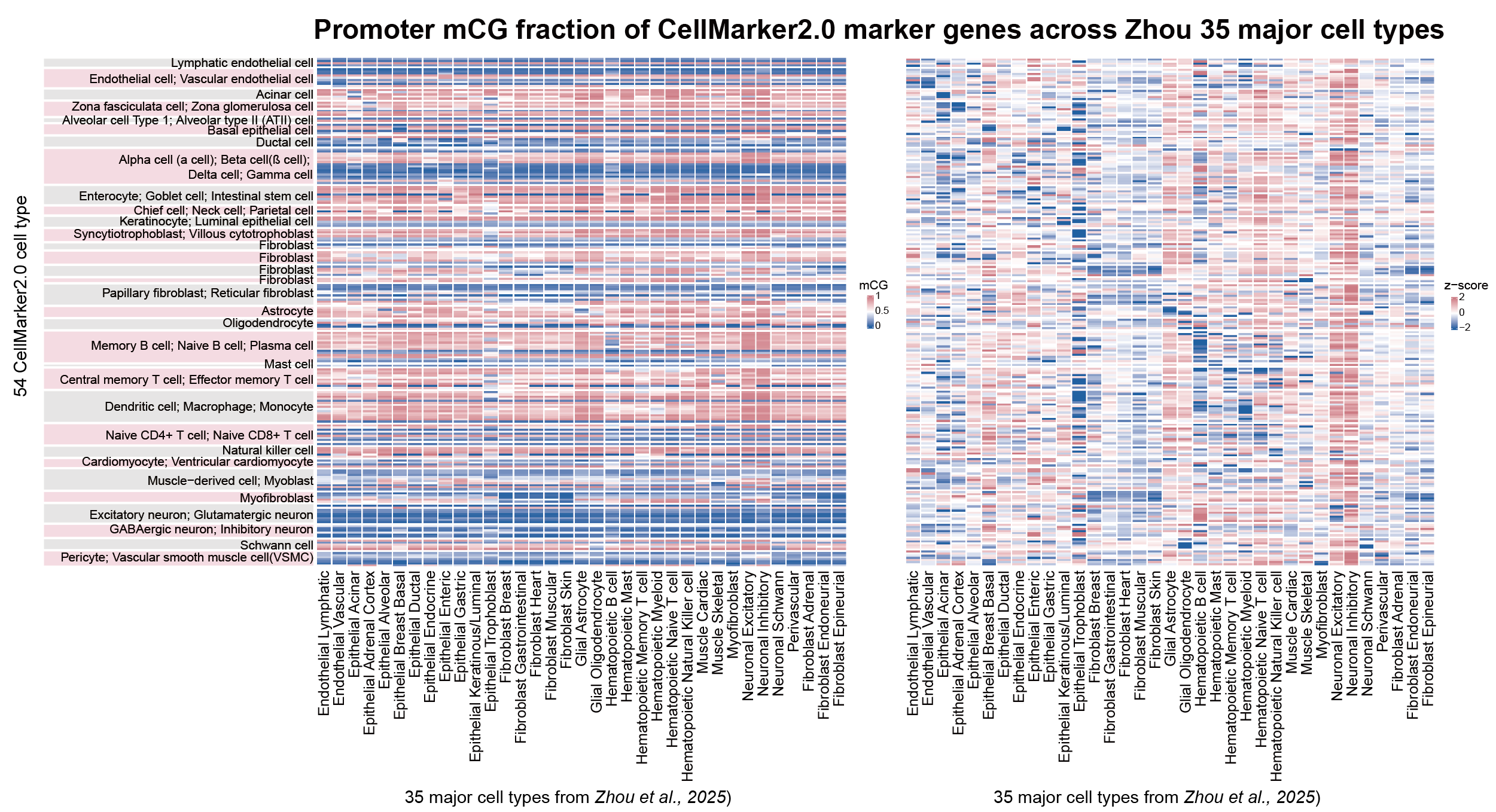
- Gene-marker control: compared with promoter methylation of CellMarker2.0 marker genes.
Results
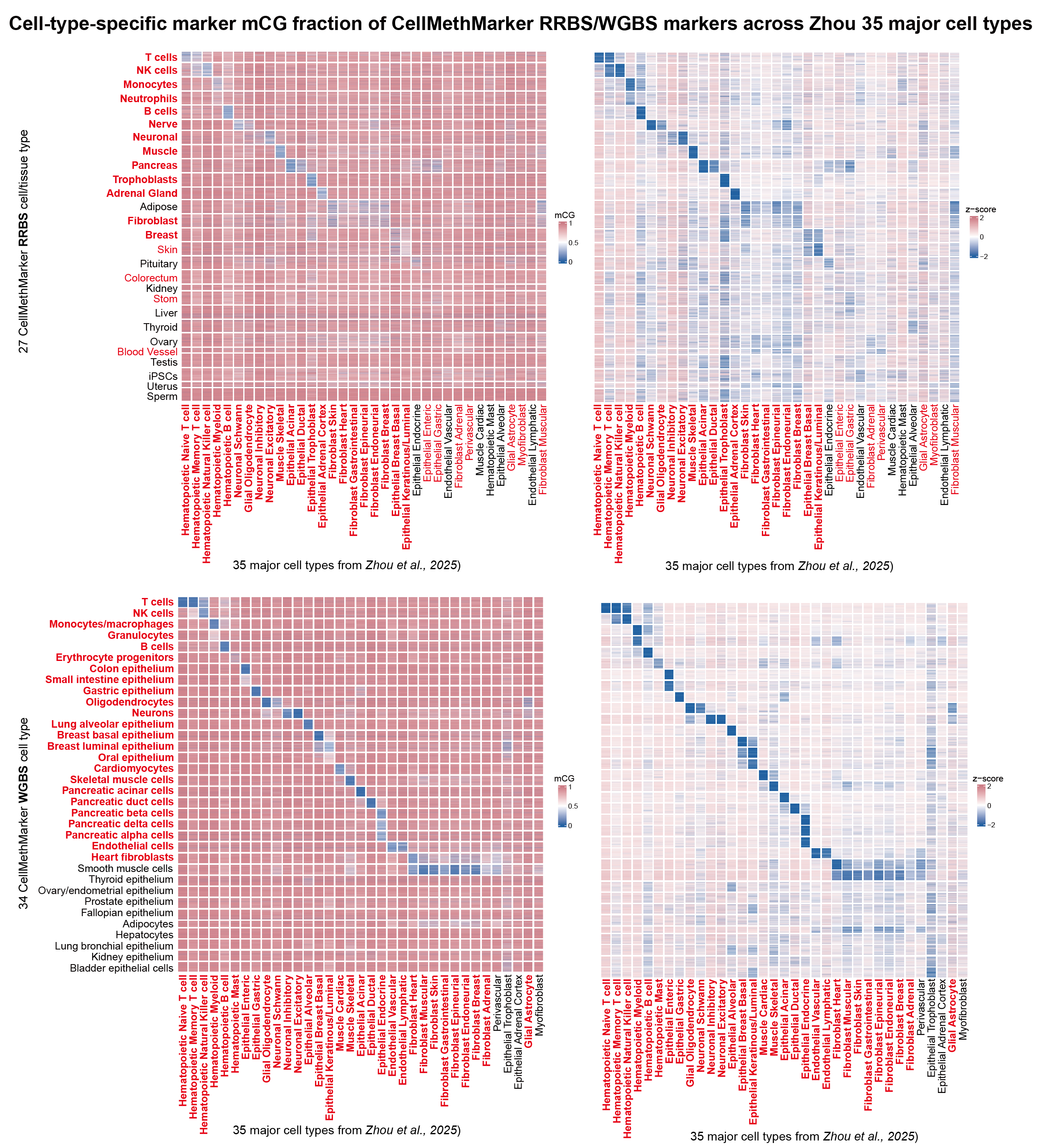
CellMethMarker markers recovered expected cell-type-specific hypomethylation patterns. WGBS custom markers showed particularly strong transferability to the independent Zhou atlas, while RRBS markers also recovered many expected low-methylation signatures.
RRBS custom markers
WGBS custom markers
Visual evidence
Marker-region methylation is more cell-type-specific than promoter methylation of conventional marker genes.
Comparison
| Feature | CellMethMarker custom markers | CellMarker2.0 marker gene promoters |
|---|---|---|
| Marker type | DNA methylation marker regions selected for cell/tissue specificity. | Conventional marker genes, evaluated here using promoter methylation. |
| Expected signal | Cell-type-specific hypomethylation at matched marker regions. | Promoter hypomethylation may or may not correspond to marker gene identity. |
| Observed specificity | Strong and interpretable low-methylation signatures, especially for WGBS markers. | Weaker and less consistent methylation specificity across major cell types. |
| Practical use | Suitable as a reference for methylation-based cluster or sample annotation. | Useful biological context, but less direct for methylation-only annotation. |
Suggested workflow for users
- Cluster single-cell methylation profiles or pseudobulk profiles.
- Profile hypomethylated marker regions or genes in each cluster.
- Compare cluster profiles with CellMethMarker marker signatures.
- Annotate candidate cell identities based on marker enrichment and heatmap patterns.
- Validate using tissue context and optional RNA/ATAC label transfer.
Notes and limitations
The label space of CellMethMarker and the Zhou atlas is not identical, so some mappings are approximate or based on biologically related categories. This analysis is intended as a practical validation of marker utility rather than a formal classifier benchmark. Future work could add quantitative enrichment scores or automated cluster-label prediction.
Reference dataset: Zhou, J. et al. Human Body Single-Cell Atlas of 3D Genome Organization and DNA Methylation. bioRxiv. https://doi.org/10.1101/2025.03.23.644697 (2025).